Facilities
µRaman Spectrometer
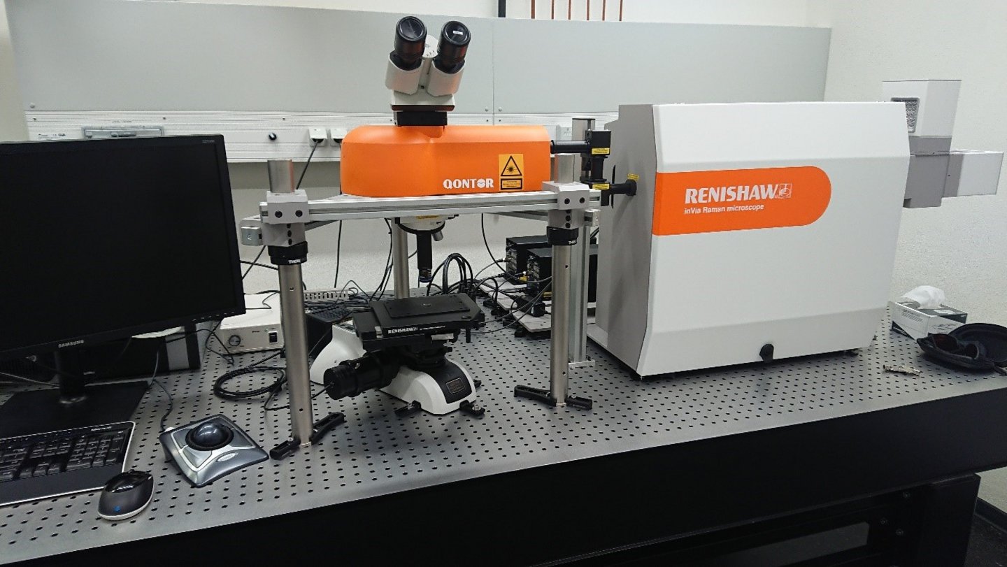
Raman spectroscopy is a non-destructive analytical technique that allows the observation of vibrational modes of (polarizable) molecular bonds. These can be used as a fingerprint to identify minerals and other substances (solid, liquid, gaseous) or characterise structural changes within the material (generated by, e.g., stress or radiation damage). With a laser beam size of about 1 µm, µRaman spectroscopy has a high lateral resolution and the laser also allows to focus inside of transparent samples for inclusion analysis. Measurements can be performed on a point-by-point basis or entire sample surfaces can be imaged fully automatically. Time-, pressure- and temperature-controlled in situ analysis can be conducted with a series of available experimental equipment, e.g. heating and cooling stages as well as diamond anvil cells.
Sample preparation for µRaman spectroscopy depends on the research question. In general, any sample (fitting under the microscope) can be analysed. Simple powders can be sufficient, however, a flat polished surface is ideal as it mitigates scattering of the laser beam.
For analytical questions or measurement time, please contact the lab manager: Dr. Christoph Lenting, e-mail: (EMAIL_FCT).
Technical details:
- 180°C backscatter geometry with microscope (10x, 100x, 50x LDW, 50x SLDW objectives), top & bottom illumination
- Laser sources: 456 nm (50 mW), 532 nm (100 mW), 786 nm (50 mW)
- Front- and back-illuminated CCD detector for precision and fast analysis
- Fully automated XYZ stage with large working space for equipment or samples
External equipment:
- Linkam heating stage TS1500 (ambient to 1500°C)
- Linkam heating-cooling stage THMS600 (-195°C to 600°C)
- Diamond Anvil Cell
- Hydrothermal Diamond Anvil Cell